
What causes reconstituted peptides to be cloudy? Common explanations include incomplete dissolution, air bubbles from vigorous mixing, aggregation or precipitation, and a mismatch between the peptide and its water, pH, concentration, or solvent. Cloudiness is an observation, not a diagnosis. When it appeared and whether it clears with gentle mixing can help distinguish temporary haze from a persistent change.
A cloudy solution can be concerning, especially if it was initially clear or contains visible particles. Appearance alone, however, cannot identify the cause or establish whether a preparation is suitable for an experiment.
Mixing technique, water quality, pH, concentration, temperature, and solvent choice can all affect clarity. The following guidance explains how to assess temporary haze, when persistent cloudiness calls for stopping and reviewing the preparation, and why visual clarity is not proof of purity, identity, or sterility.
Key Takeaways
- Use gentle mixing and allow time for dissolution; vigorous shaking can introduce bubbles and may worsen aggregation.
- Consider concentration, temperature, and freeze-thaw history when assessing persistent haze.
- Water quality and pH can affect solubility, while preservatives address a different concern than clarity.
- Select a solvent based on the compound-specific preparation method and assay requirements rather than applying a universal fix.
- Treat visible cloudiness as a signal to assess the preparation, not as a diagnosis or proof of contamination.
1. Gentle mixing and avoiding vigorous shaking
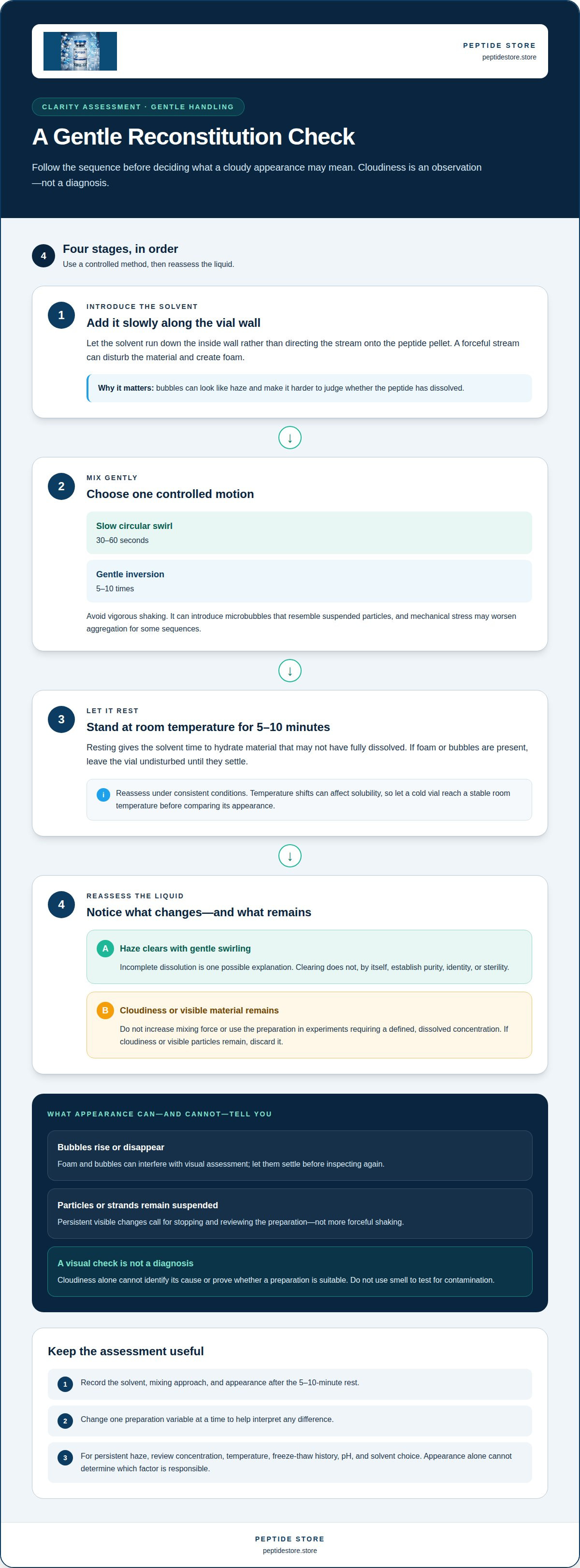
During reconstitution, the solvent needs to contact the research peptide without unnecessary mechanical stress. Add it slowly down the inside wall of the vial rather than directing the stream onto the peptide pellet. A forceful stream can disturb the material and create foam. Bubbles may then look like haze, making it harder to tell whether the peptide has dissolved.
Use a controlled sequence: add the solvent gradually, then swirl the vial in slow circles for 30-60 seconds. Gentle inversion 5-10 times is another option. Avoid vigorous shaking. It can produce microbubbles that resemble suspended particles, and mechanical forces may disrupt peptide structure. For some sequences, aggressive movement can also worsen aggregation, affecting solution clarity and stability.
- Introduce the solvent slowly along the vial wall.
- Swirl gently for 30-60 seconds, or invert the vial 5-10 times.
- Let the vial stand at room temperature for 5-10 minutes after mixing.
- Reassess the liquid before any further handling.
A brief resting period gives the solvent time to hydrate material that has not fully dissolved. If mild cloudiness clears over several minutes with gentle swirling, incomplete dissolution is one possible explanation. If the solution remains cloudy after resting, do not assume it is clear or use more forceful shaking to break up the haze.
Foam can interfere with visual assessment. If rapid solvent addition or shaking has left bubbles in the vial, let it sit undisturbed until the foam settles, then inspect it again. Note whether bubbles rise or disappear, or whether particles or strands remain suspended. If the appearance persists, stop rather than increasing the mixing force.
Gentle swirling and shaking are not equivalent. Swirling moves liquid around the vial with less agitation; shaking introduces more air and can obscure whether a research peptide has dissolved. A newly mixed vial with fine bubbles, for example, may look uniformly hazy, while slow swirling gives the material time to dissolve without as much foaming. This distinction matters before analysis because haze can complicate observations.
Keep the handling method consistent across preparations. Record the solvent addition and mixing approach, then note whether the solution clears after the 5-10-minute rest. Changing one step at a time helps identify whether mixing contributed to the appearance. Gentle technique may resolve foam or incomplete dissolution, but persistent cloudiness needs assessment rather than repeated shaking.
2. Why is my peptide cloudy after reconstitution?
Do not use a cloudy reconstituted peptide in experiments that require a defined, dissolved concentration. Allow it time to dissolve; if cloudiness or visible particles remain, discard it.
Aggregation occurs when peptide molecules associate instead of remaining evenly dispersed in solution. It depends on the molecule and its environment, including concentration, pH, solution composition, surfaces, temperature, and physical stress. A preparation may turn cloudy immediately or develop haze during storage, even if mixing initially seemed complete.
Freeze-thaw cycles can contribute to cloudiness that develops later. As ice forms and melts, the sample’s concentration changes repeatedly, which can promote aggregation. A vial that is repeatedly frozen and thawed experiences these changes with each cycle. Aliquoting a solution for storage limits repeated freeze-thaw handling of the same vial.
Concentration is another important factor. If the concentration exceeds the peptide’s solubility under the preparation conditions, some material may remain undissolved or aggregation may occur. Persistent cloudiness may indicate that the concentration is too high. Plan the target concentration before adding solvent to avoid an unsuitable preparation.
For concentration-related haze, one practical approach is to prepare a clear stock at a lower concentration, then dilute it to the working concentration. Keep the temperature stable while the peptide dissolves. If the vial is cold, let it reach a stable room temperature before reassessing its appearance. Temperature shifts can affect solubility, so compare samples only under consistent conditions.
Cloudiness alone cannot show that aggregation is the cause. Persistent particles or strands after resting, gentle mixing, and temperature stabilisation are reasons to stop and review the preparation. Aggregation and contamination can both cause visible changes, while clarity does not rule out contamination. Do not use smell as a test: it cannot establish whether a vial is contaminated.
Record the freeze-thaw history, solvent, concentration, and appearance at each assessment. Change one preparation variable at a time so that any difference is easier to interpret. Sterile water may also be relevant when cloudiness appears days after an initially clear preparation; contamination is one possible concern, distinct from concentration-driven aggregation.
For related preparation and handling context, review the peptide information alongside the compound-specific method. Keep a preparation with unexplained cloudiness separate from experiments requiring a defined dissolved concentration until it has been assessed.
3. Bacteriostatic water quality, preservative concentration, and pH
Bacteriostatic water contains 0.9% benzyl alcohol as its bacteriostatic agent. That preservative helps limit microbial growth in a multi-dose vial, but it doesn’t establish peptide solubility or prevent every cause of cloudiness. Solvent choice must still suit the compound and its preparation method.
Water quality also matters. Sterile water lacks preservatives, so microbial growth can become a concern after reconstitution. If a solution was initially clear but turns cloudy after 4-5 days, the water used is one factor to consider. Contamination can also develop without visible cloudiness. Appearance alone cannot confirm sterility or identify endotoxins; those require appropriate testing.
Concentration affects solubility. For example, adding 1 mL of bacteriostatic water to a 10 mg vial creates a concentration of 10 mg/mL. That concentration may be too high for some peptides to dissolve fully. A very small reconstitution volume can therefore leave haze or undissolved material, even when the mixing technique is gentle.
Choose a reconstitution ratio based on the compound’s preparation method and the concentration required for the research. A high-mass vial combined with little solvent creates a higher initial concentration. Peptide sequence matters too: compounds differ in how they interact with water, pH, and other solution components. A ratio that works for one sequence may not produce the same solubility in another.
pH can influence peptide charge and solubility. The reported pH range for stability of many therapeutic peptides is 4.5-7.0, but this should not be treated as a universal target for every research peptide. A peptide may become less soluble when solution conditions approach its isoelectric point. Avoid adjusting pH without a compound-specific method, since an unsuitable change can increase haze or affect the experiment.
Temperature is another preparation variable. Keep it stable during dissolution, and assess a cold solution only after it reaches a stable room temperature. Add solvent gradually and mix gently. This helps distinguish concentration or solubility problems from foam caused by handling.
If cloudiness appears days after reconstitution, review the water type, storage, vial access, and preparation timing. Sterile-water preparations can become contaminated by day 3, while a clear solution that clouds on days 4-5 warrants careful assessment. Don’t infer safety from clarity, and don’t use smell to test for contamination. Keep a preparation with unexplained cloudiness separate from work requiring a defined concentration.
4. Solvent selection, including acetic acid and DMSO
Solvent choice can affect whether a compound dissolves, remains dispersed, or forms visible haze. The right choice depends on the peptide’s properties, its compound-specific preparation method, and the assay. Sterile water is not a universal solvent for every research peptide. A liquid that dissolves one sequence may leave another cloudy or alter the sample’s behaviour.
Acetic acid may be relevant when a peptide is difficult to dissolve under neutral conditions. For some basic sequences or aggregation-prone peptides, a mildly acidic first-step solvent can improve solubility. This is not a default adjustment for every vial. The method must specify whether acidic conditions suit the compound, since pH can affect molecular charge and solubility.
DMSO may be used as a first-step stock solvent for more hydrophobic peptides. In that approach, keep the final solvent fraction consistent across samples. For example, varying the DMSO fraction between test conditions changes the solvent composition as well as the variable under study. That makes comparisons harder to interpret, even if each liquid looks clear.
Buffers and salts also affect the solution environment. Their composition can alter peptide charge, interactions between molecules, and the conditions that support solubility. A buffer suitable for one assay may not suit another. Before preparation, identify the compound-specific method and assay requirements, then follow their stated solvent and buffer conditions rather than substituting based on appearance alone.
Cloudiness after a solvent change can reflect incomplete dissolution, precipitation, or aggregation. These processes may overlap, and visual inspection cannot distinguish them with certainty. If a liquid that was initially clear becomes hazy after dilution or a pH adjustment, record the change and keep other conditions constant. Do not add acid, base, salt, or an organic solvent without a method that supports the change.
Use a straightforward selection process. First, identify the peptide and the solvent specified in its preparation method. Next, confirm that the same solvent composition is compatible with the planned assay. Then prepare the sample under controlled conditions and assess it before analysis. If the method calls for an acidic first step or a DMSO stock, follow the specified sequence and keep conditions consistent across samples.
There is no universal solvent fix for a cloudy preparation. Changing the solvent or pH can affect solubility, molecular interactions, and assay results at once. Treat a persistent change in clarity as a reason to stop and review the compound-specific method, not as a reason to keep experimenting with solvent proportions. A clear liquid alone does not establish purity, identity, or sterility.
5. Causes of cloudy peptide solutions
Cloudiness is an observation, not a diagnosis. A cloudy solution may reflect incomplete dissolution, aggregation, precipitation, or contamination. These causes can look similar in a vial but have different implications. Temporary haze, for example, may clear after the solution reaches room temperature, while persistent particles need review. Record when the change appeared and whether it followed reconstitution, storage, or vial access.
Aggregation means peptide molecules associate; precipitation means a poorly soluble phase separates from the liquid. The processes can overlap, but neither term simply means “cloudy.” A peptide may become less soluble when its isoelectric point is near the solution’s pH. Concentration, solvent composition, and physical stress also influence aggregation. A gel is different: it is a macroscopic network, not another name for a cloudy liquid.
Timing provides useful context. Haze present immediately may reflect under-dilution, a cold solution, or incomplete dissolution. Let a cold vial reach a stable room temperature before reassessing it. Cloudiness that appears after re-entering a previously clear vial can raise concern about contamination, particularly within 24-48 hours after the first re-entry. Neither timing pattern proves a cause, but both belong in the preparation record.
Use a structured comparison rather than changing several conditions at once. Record the peptide, solvent, volume, concentration, storage history, vial access, and appearance. Compare those details with the compound-specific preparation reference. To assess a possible technique issue, keep the temperature stable and use only the mixing steps specified by the method. A clearer sample after one controlled change helps narrow the possibilities; it does not establish identity or sterility.
If persistent cloudiness is identified early, additional diluent may be considered only when the compound-specific method supports it. Do not add solvent arbitrarily: changing volume also changes concentration and may affect later analysis. One preparation guide uses 50% more bacteriostatic water as a reassessment point, and advises discarding the vial if cloudiness remains. That is not a universal adjustment for every peptide or assay.
Visible particles or strands that remain after resting, gentle mixing, and temperature stabilisation are reasons to stop and review. Keep the preparation separate from experiments that require a defined dissolved concentration. Do not smell the vial to assess contamination, and do not assume clarity rules it out. A clear solution can still be contaminated, while cloudiness alone cannot identify microbes or aggregation.
The practical takeaway is to interpret appearance alongside timing, volume, storage, and handling records. Preserve the original observations before making a supported change, then document the result. If cloudiness persists, particles remain, or the solution changes unexpectedly, withhold it from concentration-dependent analysis until it has been assessed. No visual sign can replace a compound-specific method or appropriate testing.
Contamination is one possible explanation for cloudiness, especially when haze develops after a previously clear vial is accessed again. This timing raises concern but does not prove the cause.
Peptide Store
Peptide Store supplies research-grade compounds for laboratory and scientific applications. After identifying how solvent choice, concentration, and handling can affect clarity, researchers can use its catalog to source compounds such as Tirzepatide, Retatrutide, Semaglutide, and Cagrilintide for research purposes. A cloudy preparation still requires assessment against the relevant compound-specific method; sourcing a peptide does not diagnose or resolve changes in a reconstituted solution.
For researchers selecting a compound for a laboratory project, Peptide Store provides a relevant next step. Contact Peptide Store with questions about its research-grade peptide offerings.
Build a More Reproducible Preparation Record
Treat an unexplained change in appearance as a prompt to strengthen preparation records. Recording the compound, preparation conditions, storage history, and observed changes gives future experiments a clearer basis for comparison. These records can also help distinguish a one-time handling issue from a recurring pattern across preparations.
Understanding what causes reconstituted peptides to be cloudy is only one part of reliable laboratory work. Apply compound-specific methods consistently and keep observations linked to each sample. This supports clearer interpretation when results differ and helps prevent visual appearance from standing in for analytical evidence.
For research involving peptide compounds, select materials that fit the project and its documented methods. Peptide Store offers Tirzepatide, Retatrutide, and Semaglutide for research applications. Discuss your research requirements with Peptide Store as you plan your next study. A careful, repeatable approach gives each preparation a stronger foundation for meaningful analysis.
Frequently Asked Questions
Why is my peptide cloudy after reconstitution?
To assess what causes reconstituted peptides to be cloudy, consider whether the haze appeared during preparation or later. Incomplete dissolution, aggregation, precipitation, or contamination can all change a solution’s appearance. If cloudiness develops after vial access, for example, record that event alongside storage conditions. Appearance alone cannot establish the cause, so keep an unexplained preparation separate from work requiring a known concentration.
Cloudiness, precipitate, gel or foam: what is different?
Foam consists of bubbles that may rise or dissipate, while precipitate is a separate, poorly soluble phase. Aggregation describes peptide molecules associating and can overlap with precipitation. A gel is a macroscopic network, not simply a cloudy liquid. Note whether the appearance moves, settles, forms strands, or holds its shape. These observations help describe the sample but do not identify its composition.
Does a clear solution prove purity or sterility?
No. Clarity cannot establish a peptide’s purity, identity, concentration, or sterility. A contaminated vial can appear clear, and visual inspection cannot detect every dissolved impurity. A transparent sample may still have an uncertain storage or handling history, for example. Use analytical methods appropriate to the research question, and treat appearance as one observation rather than a substitute for testing.
What about cloudy retatrutide?
Assess cloudiness in retatrutide using the same core distinctions as for any research peptide: incomplete dissolution, aggregation, precipitation, or contamination. Do not assume the appearance is a known feature of the compound. Compare the preparation with its compound-specific method, and withhold a sample with unexplained particles or persistent haze from concentration-dependent experiments until it has been assessed.
Why Does Reconstitution Matter So Much?
Reconstitution determines the solution’s composition and concentration, both of which influence how results can be interpreted. If samples use different solvent volumes or preparation conditions, their outcomes may not be directly comparable. A consistent, documented method links each observation to its preparation history. That record is especially useful when repeating an assay or investigating an unexpected result.
How do peptide sequence and hydrophobicity affect solubility after reconstitution?
Peptide sequences differ in their chemical properties, so they interact differently with water and other solvents. Hydrophobic sequences may dissolve poorly in aqueous conditions, while other sequences respond more strongly to pH or solution composition. A solvent suitable for one compound, for example, may not work for another. Follow the compound-specific method and keep solvent conditions consistent across assay samples.
How do pH, salt concentration, and buffer choice cause peptide precipitation?
To understand what causes reconstituted peptides to be cloudy, consider how pH, salt levels, and buffer composition affect peptide charge and molecular interactions. Solubility may decrease under conditions unfavorable to a sequence, allowing material to separate from solution. A buffer suitable for one peptide or assay may not suit another. Use the specified preparation conditions, and don’t adjust pH or salt concentration without a method supporting the change.
How can bacterial contamination or endotoxins make a reconstituted peptide cloudy?
Contamination can cause visible changes, but cloudiness alone cannot establish that bacteria or endotoxins are present. Appearance cannot identify endotoxins; appropriate testing is required.


